
Have you ever looked at the world
& felt that it was upside down?
You felt like something was OFF or just didn't make any logical sense the way it was done, built, designed, run, or created?
Do you know how Bright Field microscopy works?
It's simply shining a light from below onto a glass slide while we view the same slide from above. Because we are using such thin sections to view (usually a drop or two flattened between two pieces of glass: a cover slip and a slide), we can see through the microbes and see their insides even!!
BUT for ROOTS: they are THICK, so folks tend to slice them up or crush them to view them, and even then we are looking at silhouettes.
A Silhouette is NEVER an accurate representation of
a three dimensional object.

The Story of Manual Lighting
I just felt like it was strange - Why is the light only linear? I can't tilt things, I can't manipulate the light enough... I should give you some context: my roommate in college was a photography major. One thing I picked up from him about image creation was LIGHTING is EVERYTHING, so I do things differently from everyone I've ever seen with a microscope. I turn off the lights in the room. I don't use the eyepieces - I don't want to damage my eyes, and I like to work for extended periods of time, and I also prefer to show what I'm doing to others and to capture the images at the highest level of resolution I can because clarity is the key to identifying anything properly with a microscope, so I use a 4k camera and a 4k screen. I see what the images will look like as I record them. It's also so much more enjoyable and easier to use, and if you are following the progress of labs worldwide, they are all getting attached screens over eyepieces - it's not the future, it's already here.
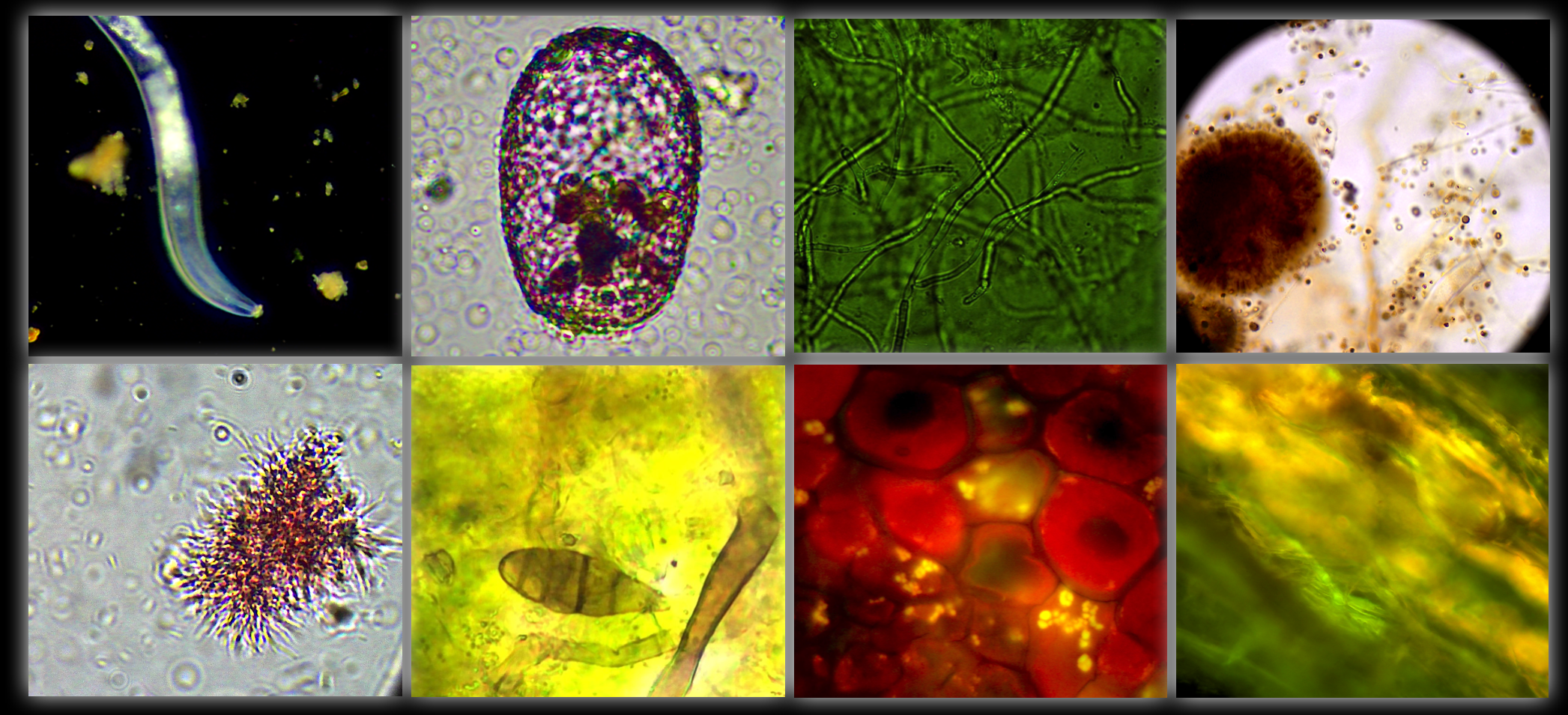
SO, I'm playing with the lights - I already have a dark field condenser (which is dispersed indirect lighting - it makes things look like deep interstellar space or the bottom of the ocean) and epifluorescence lighting (a targeted wavelength of excitation that reveals fungi and phosphorus visually - it glows NEON GREEN), and I'm doing things like turning off the base microscope entirely, boosting brightness compensation to explore all the capabilities of the microscope... BUT THEN...
I tried something novel - something I can teach people everywhere. I figured out there is an angle at which the roots will LIGHT UP, and it's not hard.
LOOK AT WHAT IT REVEALS:


What ARE We SEEING??
This is what the root looks like in low lighting - it's clear. In fact. all the plants I've ever visualized with epifluorescence and manual lighting have appeared as if the cells are made of glass. This makes sense when we think about how a greenhouse traps heat and energy - plants are absorbing light, performing photosynthesis, and even shining light out of their roots in the soil. That last one (the shining roots) didn't make any sense to me until I started working with different lighting. Once I saw that it was like glass and the soil clay, sand, and silt are all like bits of broken glass too (they're all silicates!!), I realized, that light was being conducted and refracted through organisms both to capture the energy and to allow it to conduct further down into the soil profile.
Both epifluorescence and manual lighting are light from above. That's why they appear this way.

Why is this Important?
We're seeing the roots and leaves for the first time all over again as we change the lighting, understand more of what each new image means, and map out further the new regenerative science of plants, soil, and microbes.
We can see a color difference in the meristem (tip) cells of the root and we can see the root itself more clearly than any other method that I've ever encountered.
We can see the root as a three dimensional object: it's not a silhouette or something you are blasting with light and distorting the image. We can examine the root hairs in real-time on top and at angles which allows us to diagnose how effectively the plant is performing rhizophagy (root hairs are a result of this process).
We can see things as they really are. This may be the most important thing. We need tests that show us as close to reality as possible. These roots are not stained. They aren't drowned in water. The minerals are still sticking to their root hairs. They aren't cut or crushed between glass. These are roots that are literally placed below the microscope lens on a single glass slide.
It's just that easy.
It's the fastest and easiest method EVER!!
But what's more is it is also the most natural, freshest, and therefore the most accurate way of imaging our roots. And it all started with a question:
What's the most natural in this situation?
And that has been the guiding principle in my microscopy work. That is what is at the core of the new book: https://www.thepermaculturestudent.com/shop/regenerative-soil-microscopy

